
Dr. Uthai Rutnin was the first ophthalmologist to use an Indirect Ophthalmoscope to examine the retina and perform modern retinal surgery.

The first in Thailand to acquire the Ruby Laser, the first ‘true’ laser developed for the eye. At the time, we also introduced the use of a conjunctival auto-graft upon the removal of a pterygium (benign conjuntival mass), thus lowering the rate of recurrence to 5-10% as compared to 40-50% without a graft.

The first in Thailand to use the Argon Laser to treat glaucoma, diabetes and retinal disease.

One of the first hospitals in Thailand performing phaco-emulsification (ultrasound treatment of cataracts).

One of the first hospitals in Thailand using the Excimer laser to correct vision problems (myopia, hyperopia and astigmatism).

The first private hospital in Thailand to use coral implants for artificial eye providing more natural movement than the silicone or glass implants.

The first in Thailand to offer Implantable Intraocular Lens for the treatment of high degrees of short-sightedness, incurable by LASIK treatment.

Deep anterior lamellar keratoplasty (DALK) is a surgical procedure for removing the corneal stroma down to Descemet’s membrane. It is most useful for the treatment of corneal disease in the setting of a normally functioning endothelium.

The first in Thailand to use Optical Coherent Topography, the most up-to-date method for retina analysis.

One of the first hospitals in Thailand performing phaco-emulsification with Premium IOLs provide both distance and near focus at the same time. The lens has different zones set at different powers.

The first private hospital in Thailand to use Endoscopic DCR to perform surgery on blocked tear ducts without sutures being required.

The first group of hospitals in Thailand to use Multi-focal intraocular lenses, allowing patients to focus near-by and far away after cataract surgery without the use of glasses.

The first in Thailand to use the Ferrara ring to treat corneal keratoconus.

The first in Thailand to use HRA-2 technology to diagnose and photograph diseases of the retina.
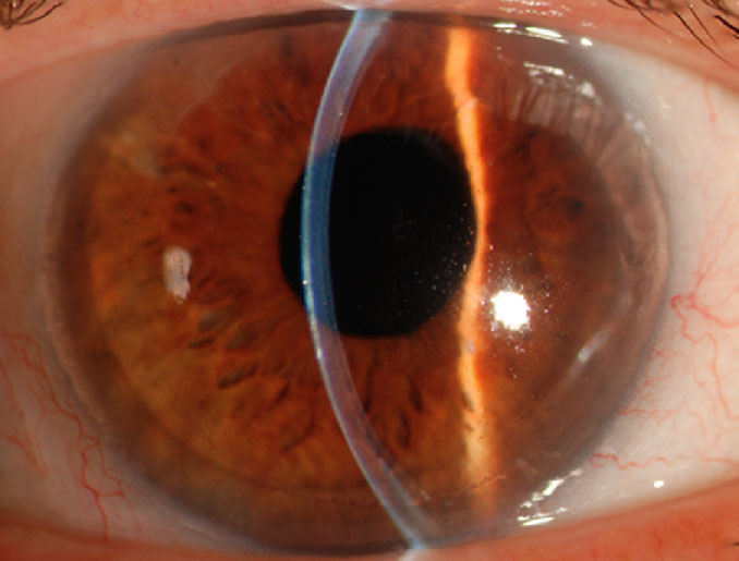
Descemet's Stripping Automated Endothelial transplantation (DSAEK) was performed. DSAEK is the posterior lamellar corneal transplant surgery. Only 25-30 percents of total corneal thickness is transplanted. The surgical wound size is 5 millimeters which is smaller than the conventional (penetrating keratoplasty) surgery. The surgery has stronger wound and faster recovery after the surgery than the conventional surgery.
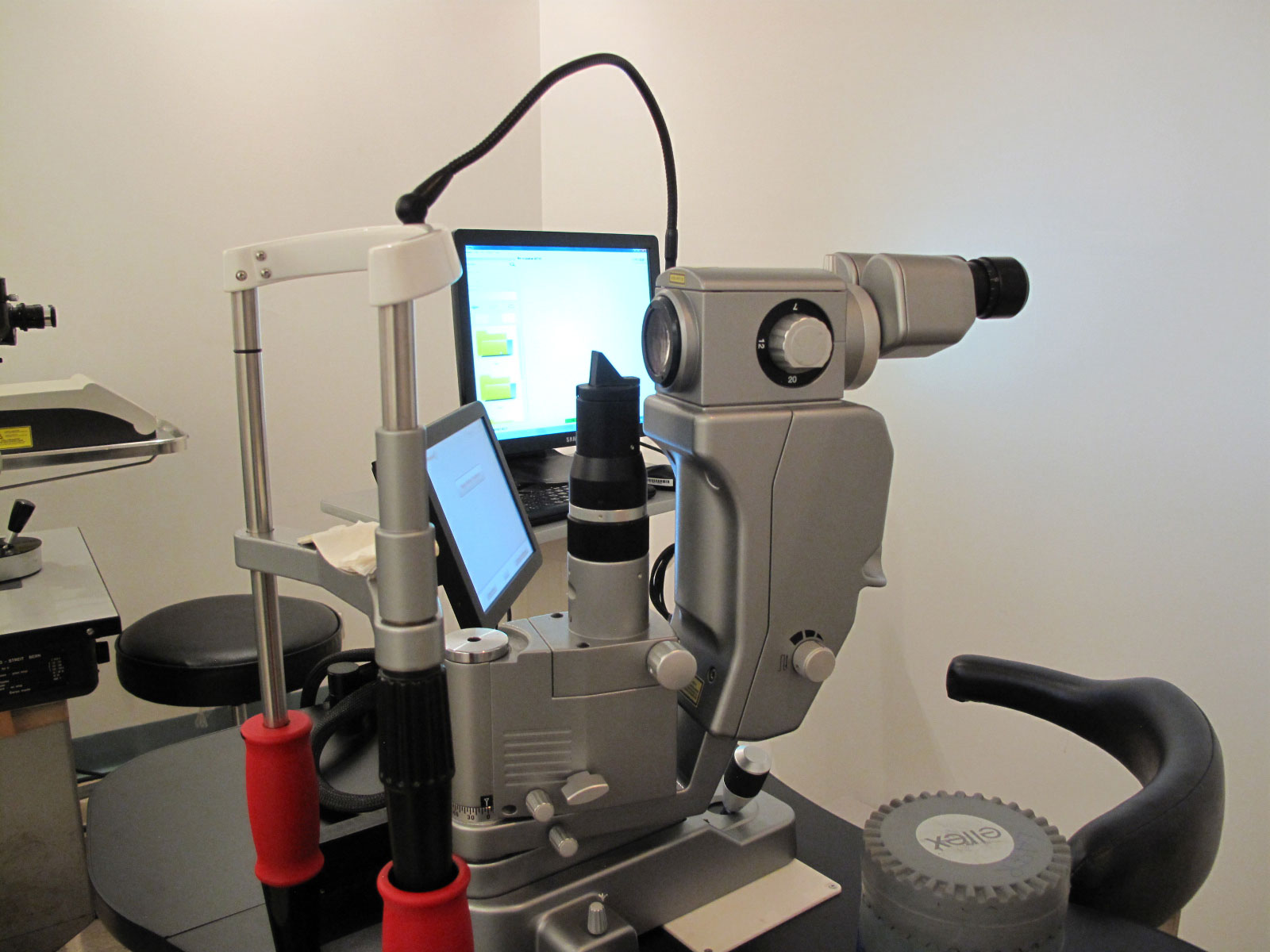
The first hospital in Thailand to use pattern scan laser technology (PASCAL).
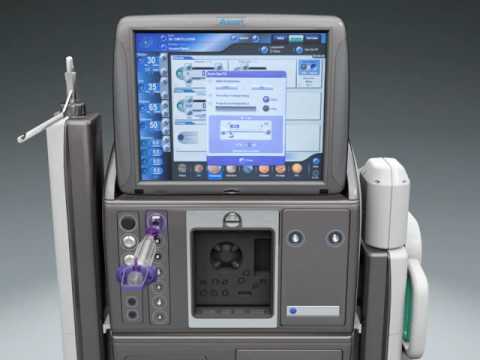
The first hospital in Thailand to use the Constellation system for vitrectomy and retinal surgery.

Descemet's membrane endothelial transplantation (DMEK) surgery was first successfully performed in Thailand. DMEK is a selective lamellar corneal transplant surgery which only Descemet's membrane and corneal endothelial cells of 10-20 microns or 10 percents of total corneal thickness was transplanted. The surgical wound size is only three millimetres. Recovery period takes about one month only. It is reported from the study that the rejection rate of DMEK is 15-20 times less than the conventional (penetrating keratoplasty) and Descemet's Stripping Automated Endothelial transplantation (DSAEK). DMEK surgery is currently the surgery of choice in patient with corneal decompensation.

The first Eye Hospital who bring the new retina camera (OPTOS) that use laser instead of regular light that scan the retina to study the blood flow of the retina. It can take the picture of the retina as wide as 200 ° when normal retina camera can take only 50 ° This can be done without dilating the pupil, but it will give better detail if the pupil is dilated. Because if use a laser light , the doctor can study the deeper layer "choroid", which the older model can see only superficial layer "retina". It is a much improved technology.
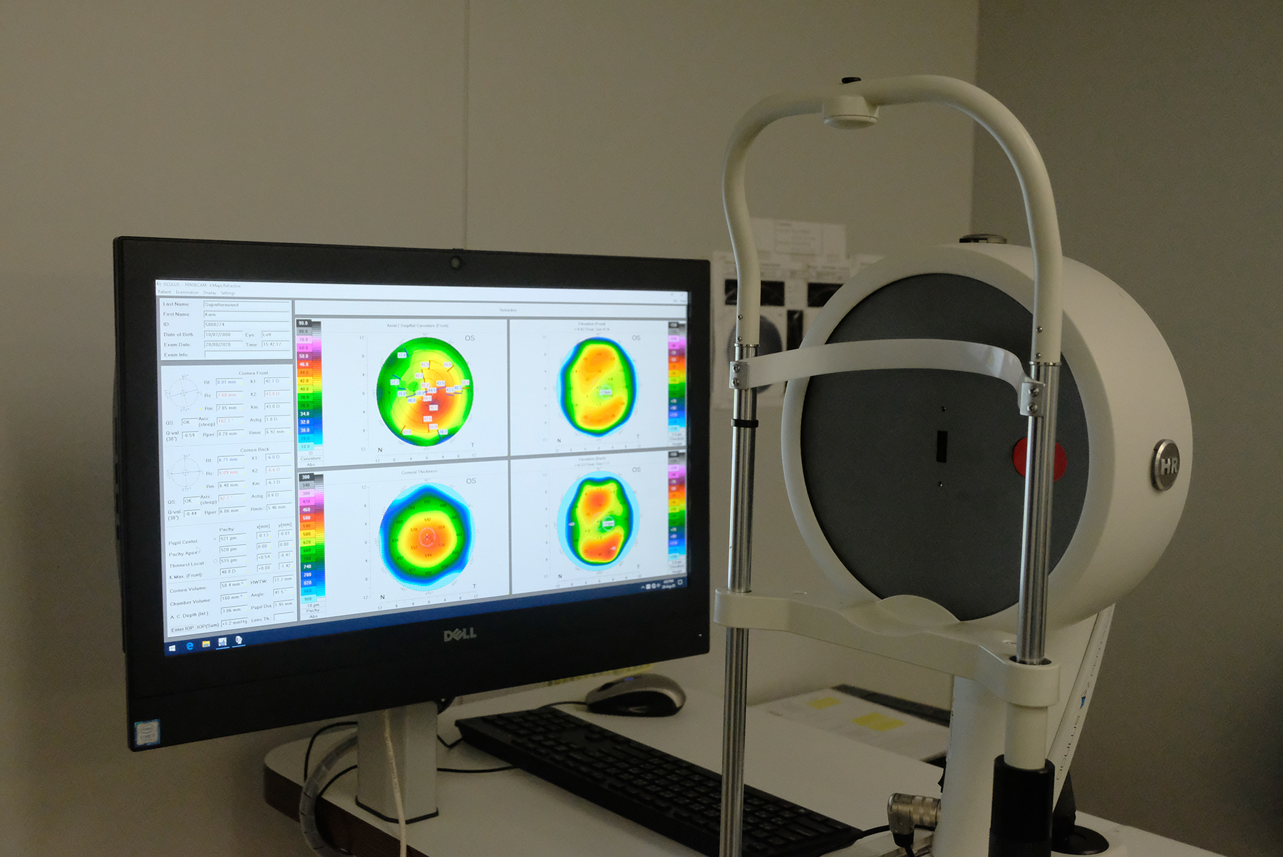
Corneal tomography system. It creates a 3-D imaging of the anterior segment and provides details of the anterior and posterior corneal contour, pachymetery, anterior chamber depth and pupil diameter. This makes it easier to diagnose patient who requires corneal transplant or cataract surgery with Toric Lens. The pentacam will provide more accurate test result.
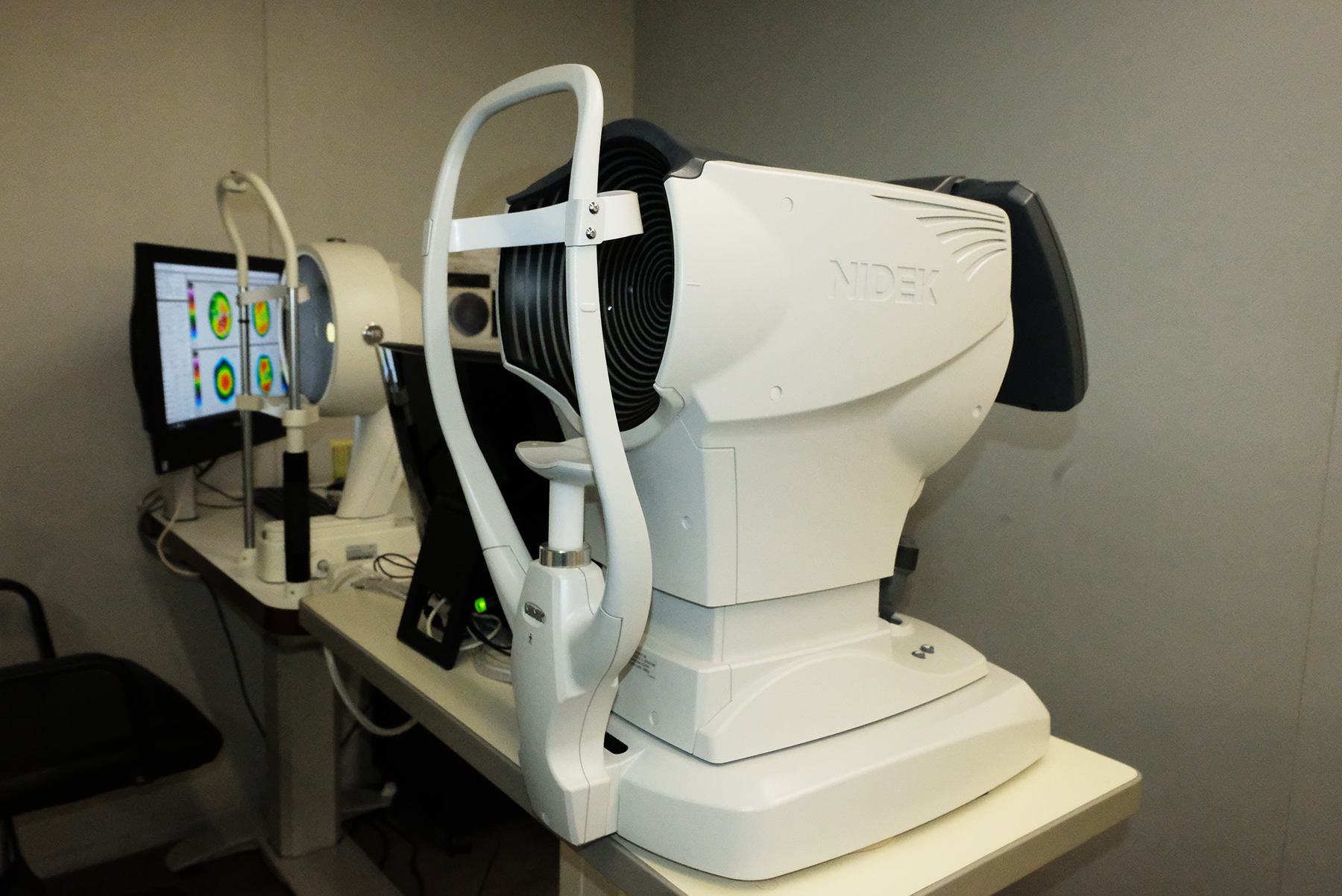
OPD Scan provides anterior segment and corneal surface analysis for corneal diseases such as keratoconus.
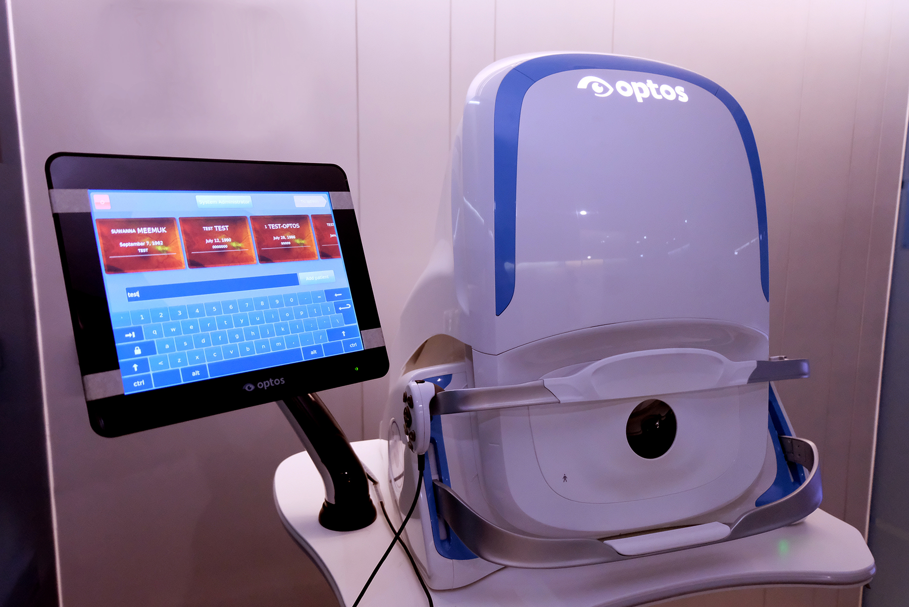
The imaging machine which analyze the blood circulation system in the retina. It produces a 200°, single-capture retinal image of unrivaled clarity in less than ½ second and is changing management of diseases including DR, AMD, and uveitis.
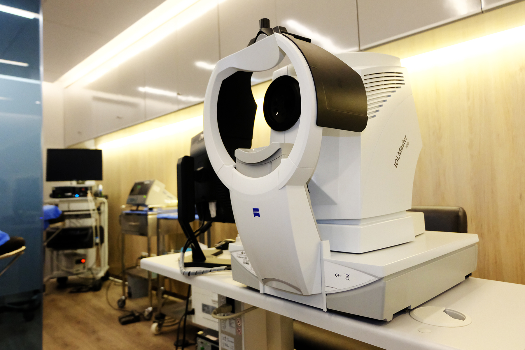
The IOLMaster 700 is the latest technology of non-contact intraocular lens measuring device for Cataract Surgery. Intended to reduce the risk of refractive surprises, this device provides highly repeatable and accurate lens thickness, anterior chamber depth, and corneal thickness measurements of the eye.
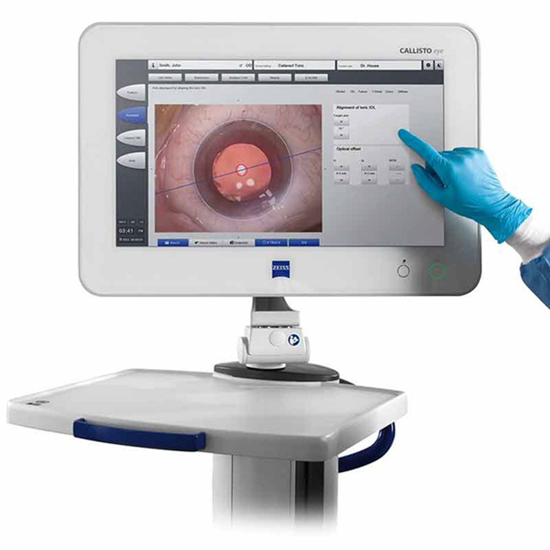
CALLISTO is a technology designed to work together seamlessly to achieve excellent results in cataract surgery, including markerless toric IOL alignment. With welding system connect the data from the IOLMaster 700 to bring the measured value to display to the ophthalmologist during the surgery in real time.