 02-056-3333

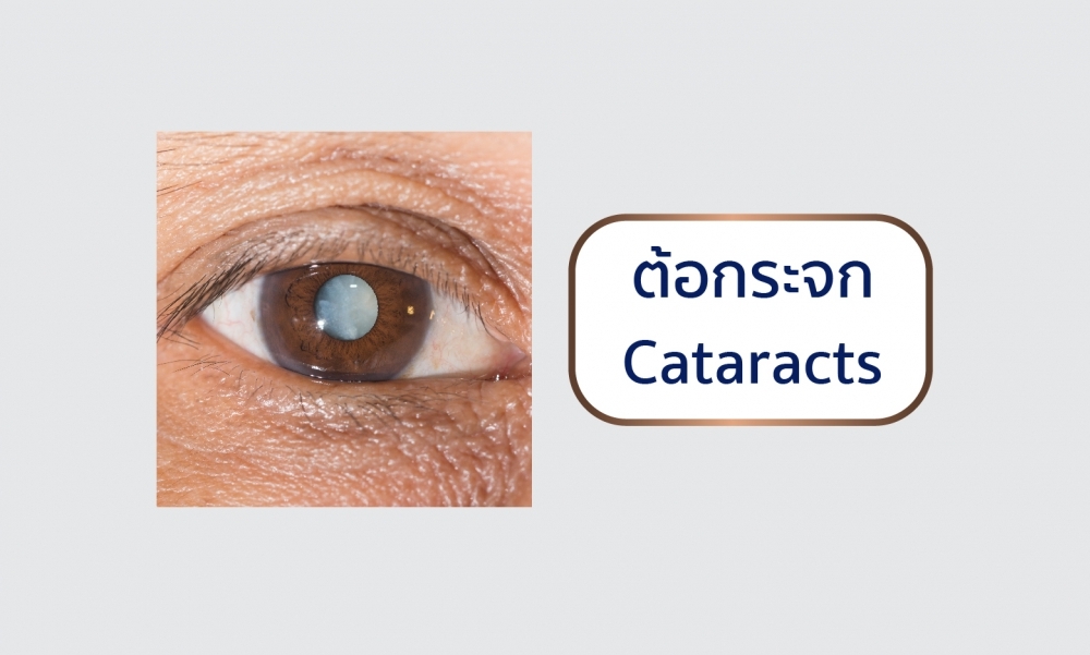
Cataract is a clouding of the eye's lens. When we look at something, light rays travel into our eye through the pupil and are focused through the lens onto the retina, a layer of light-sensitive cells at the back of the eye. The lens must be clear in order to focus light properly onto the retina. If the lens has become cloudy, this is called a cataract.
Cataract Symptoms
Most age-related cataracts develop gradually. As a result, you may not notice signs or changes in your vision right away when cataracts first develop.
Cataract symptom progression
- Painless cloudy, blurry or dim vision
- More difficulty seeing at night or in low light
- Sensitivity to light and glare
- Seeing halos around lights
- Faded or yellowed colors
- The need for brighter light for reading and other activities
- Frequent changes in eyeglass or contact lens prescription
- Double vision within one eye
Cataract as part of aging
The lens is made mostly of water and protein. As we age, the lens continues to grow layers on its surface and hardens. Protein in the lens may clump together and become cloudy in some areas, preventing light from passing clearly through the eye.
Congenital or developmental cataracts
This type of cataract can occur in infants or children. They may be hereditary or they can be associated with some birth defects. Some occur without any obvious cause.
Non-age related cataracts from other disease or medication
These cataracts are caused by other eye diseases or previous eye surgery. Chronic disease can make you more likely to develop cataracts; for example, diabetes has been proven to increase risk for cataracts. Excessive use ofsteroid medications can spur development of this type of cataract as well.
Traumatic cataracts
These cataracts are related directly to an eye injury. Traumatic cataracts may appear immediately following injury, or they can develop several months or even years later.
Cataract Diagnosis
During a comprehensive, dilated eye exam (where your pupil is widened with eye drops), your Eye M.D. will examine and test your eyes to make a cataract diagnosis.
Once I have a cataract diagnosis, what should I do?
- Have an eye exam every year if you're older than 65, or every two years if younger.
- Protect your eyes from UV light by wearing sunglasses that block at least 99 percent UV and a hat.
- If you smoke, quit. Smoking is a key risk factor for cataracts.
- Use brighter lights for reading and other activities; a magnifying glass may be useful, too.
- Limit driving at night once night vision, halos or glare become problems.
- Take care of any other health problems, especiallydiabetes.
- Get the right eyeglasses or contact lenses to correct your vision; when it becomes too difficult to complete your regular activities, consider cataract surgery.
- To make an informed decision about cataract surgery, thoroughly discuss with your ophthalmologist the surgical procedure, preparation for and recovery after surgery, benefits and possible complications of cataract surgery, cataract surgery costs, and other important information.
- Do not use eye drops or other treatments that claim to dissolve or remove cataracts. There is no proven way to dissolve cataracts with eye drops. Surgery is the only way to remove cataracts
Cataract Treatment
If your vision is only slightly blurry, a change in your eyeglass prescription may be all you need for a while. However, after changing your eyeglass prescription, if you are still not able to see well enough to do the things you like or need to do, you should consider cataract surgery.
With cataract surgery, your eye's cloudy natural lens is removed and replaced with a clear artificial lens implant (called an intraocular lens, or IOL). Your ophthalmologist will discuss the cataract surgery procedure, preparation for and recovery after surgery, the benefits and possible complications of cataract surgery, cataract surgery costs and other important information.
Cataract surgery is often performed as an outpatient procedure and does not require an overnight hospital stay.
The cataract surgery procedure
The most common procedure used for removing cataracts is called phacoemulsification. A small incision is made in the side of the cornea (the front part of your eye), where your Eye M.D. inserts a tiny instrument that uses high-frequency ultrasound to break up the center of the cloudy lens and carefully suction it out.
After the cloudy lens has been removed, the surgeon will replace it with an intraocular lens (IOL)
Implant made of plastic, silicone or acrylic. This new, clear lens allows light to pass through and focus properly on the retina. The IOL becomes a permanent part of your eye. In most cases, the IOL is inserted behind the iris, the colored part of your eye, and is called a posterior chamber lens. Sometimes, the IOL must be placed in front of the iris. This is called an anterior chamber lens. When the IOL is in place, the surgeon closes the incision. Stitches may or may not be used. After the surgery, your Eye M.D. usually places a protective shield over your eye.
You will spend a short period of time resting in the outpatient recovery area before you are ready to go home. Following your surgery, it is very important to put in the eye drops exactly as prescribed by your ophthalmologist to promote healing. You will also need to take care to protect your eye by wearing the eye shield whenever you sleep, and by wearing special wraparound sunglasses in bright light. Be sure not to rub your eye.
During the first week of your recovery, you must avoid strenuous activity such as exercise or bending and heavy lifting (including anything over 25 pounds). You will also need to avoid getting any water, dirt or dust in your eye, which can lead to infection.
You may have some blurry vision a few days to weeks after surgery procedure. If you experience any pain or loss of vision, be sure to call your ophthalmologist.
by www.aao.org
